1. Canine Myasthenia Gravis
Perhaps the most common disease of the neuromuscular junction in dogs is canine myasthenia gravis. This disease appears to be similar in its pathophysiology to the human form of the disease. It is caused by a breakdown of the transmission of impulses from the nerves to the muscles. This keeps the muscles from contracting, causing affected dogs to show signs of weakness and fatigue – the primary symptoms of this neuromuscular disease.
Though rare, myasthenia gravis can be an inherited congenital defect that pups are born with. The congenital disorder (present at birth) becomes apparent at 6-8 weeks of age. Some breeds at risk for the congenital form are Jack Russell terriers, Springer spaniels, and Smooth fox terriers. Smooth-haired miniature dachshunds have an autosomal recessive mode of inheritance for the disease.
More commonly, myasthenia gravis is an acquired problem in adult dogs that has a bimodal age of onset – either at 1-4 years of age, or 9-13 years of age. It is thought to be the result of a defect in the dog’s immune system caused by antibodies that mistake the dog’s muscle receptors as “non-friendly,” and attack them. Multiple factors are involved, including environmental, infectious, and hormonal influences.
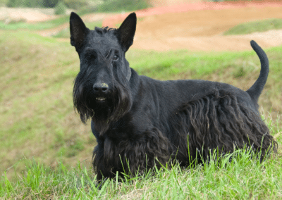
Although all breeds, male and female, are at risk the acquired form is more notable in golden retrievers, German shepherds, Labrador retrievers, Dachshunds, Scottish terriers, and Akitas. The familial form of acquired myasthenia gravis occurs primarily in the Newfoundland and Great Dane breeds. Dogs that acquire mild forms of this disease later in life have a fairly good prognosis, so long as they receive proper, timely treatment.
Risk factors include:
- Appropriate genetic background
- Tumor or cancer – particularly thymus tumor
- Immune-mediated disease
- Vaccination can exacerbate active myasthenia gravis
- Intact (non-neutered) female
Symptoms
Symptoms of myasthenia gravis can vary greatly from dog to dog. The most common symptom is muscle weakness that worsens with exercise but improves with rest. Clinical presentation can include localized involvement of the muscles of the throat and esophagus (trouble drinking and swallowing), the muscles adjacent to the eye, and acute generalized collapse.
Any dog with acquired enlargement of the esophagus, loss of normal reflexes, or a mass in the front central area of the chest should be evaluated for myasthenia gravis. Regurgitation is common, so it is important to differentiate this from vomiting.
Signs to look for:
- Voice change
- Exercise-related weakness
- Progressive weakness
- Fatigue or cramping with mild exercise
- Acute collapse
- Decreased or absent blink reflex
- Sleeps with eyes open
- Excessive drooling, repeated attempts at swallowing
- Absent gag reflex
- Difficulty breathing with aspiration pneumonia
Diagnosis
There are other neuromuscular disorders, such as tick paralysis, that may have similar symptoms, so your veterinarian will want to rule them out before coming to a definitive diagnosis. To do that, he will need a careful history, a thorough physical and neurologic examination, and specialized laboratory testing.
A complete blood profile will be conducted, including a chemical blood profile, a complete blood count, and a urinalysis. Your veterinarian may also check for such things as thyroid functioning. Diagnostic imaging will include chest X-rays to look for an enlarged esophagus and aspiration pneumonia, and an ultrasound-guided exploration of the chest, to look for a mass. If a mass is found, a biopsy will need to be performed to confirm whether the growth is cancerous. Additional tests may include a myelogram (contrast x-ray of the spine) and a CT or MRI of the spine and brain. They may also conduct a Tensilon test, which checks muscle response.
The diagnosis of myasthenia can be confirmed either pharmacologically or electro-physiologically. The pharmacologic test depends upon the animal’s response to a short-acting acetylcholinesterase inhibitor. Marked improvement in strength in response to the slow intravenous injection of less than 2.2 mg/kg of edrophonium chloride is considered positive for a clinical diagnosis of myasthenia. Neuromuscular junction testing of myasthenic animals demonstrates a decremental response of 20% or more upon repetitive stimulation at rates less than 7 Hz. This decrement can be eliminated through the use of cholinesterase inhibitors.
Treatment
Your dog will need to be hospitalized until adequate dosages of an anticholinesterase drug are determined. Treatment consists of an oral dosage with long-acting anticholinesterases such as neostigmine bromide. In the dog, this medication can usually be eliminated after less than 2 to 3 weeks.
Because dogs with myasthenia gravis have a poorly functioning esophagus, they need to eat or be fed carefully. Make sure that your dog’s head is elevated during feeding and for 10—15 minutes afterward. Pneumonia can be a very serious side effect of any disorder that impacts a dog’s ability to swallow correctly. If your dog has aspiration pneumonia, it may require intensive care in a hospital setting. Nutritional maintenance with a feeding tube and multiple feedings of a high-caloric diet will be necessary if the dog is unable to eat or drink without significant regurgitation. Oxygen therapy, intensive antibiotic therapy, intravenous fluid therapy, and supportive care are generally required for aspiration pneumonia. If a tumor is found during exploration, surgery will be required.
Management
You should see a return of muscle strength once the appropriate treatment has been found. Your veterinarian will want to perform chest X-rays every 4-6 weeks for resolution of the enlarged esophagus. Your doctor will also do follow-up blood tests every 6-8 weeks until your dog’s antibodies have decreased to normal ranges.
Prevention
Unfortunately, there is no prevention or cure for this disease. Treatment and vigilant at-home care can help dogs with this disease maintain a quality life for a long time. The more attention paid to the prevention of aspiration pneumonia, the better the prognosis.
2. Tick Paralysis
In certain areas of the United States and in other parts of the world, a common disease of the neuromuscular junction is that produced by a toxin present in the saliva of gravid ticks of many species. This disease, tick paralysis, is seen as an ascending paralysis of skeletal muscle produced by the inhibitory effects of the toxin on the presynaptic release of acetylcholine in the nerve terminal. Clinical signs include weakness, which may be profound, without sensory deficit. The disease may be fatal, with death due to respiratory paralysis. In the United States, recovery from paralysis usually occurs within 1 to 3 days after removal of the ticks.

3. Polymyositis
The most frequently seen non-traumatic disease of muscle in the dog is Polymyositis. The primary cause of Polymyositis is an idiopathic immunologic attack on muscle tissue resulting in inflammation primarily involving skeletal muscles. Additional causes include immune —mediated infections, drugs and cancer. Various breeds of dog, including the Newfoundland and Boxer, may be affected by Polymyositis.
Dermatomyositis is a form of Polymyositis in which characteristic skin lesions are also present. Breeds most susceptible to Dermatomyositis are Rough-coated collies, Shetland sheepdogs, and Australian cattle dogs.
Symptoms
This condition produces a variety of signs that may be either acute or insidious and chronic in nature. Polymyositis produces a spectrum of presenting signs. Affected animals may manifest only certain aspects of the disease, and any animal showing lameness or painful muscles for which other causes are not obvious should be suspected of Polymyositis.

The most common sign and the most easily recognized is trismus (lockjaw), which persists even under anesthesia. When trismus is present, it may be accompanied by either swelling or
- Stiff-stilted gait
- Stiff, painful neck
- Muscle swelling
- Muscle weakness
- Muscle pain (especially when muscles are touched)
- Exercise intolerance
- Occasionally the muscles of the larynx and pharynx causing problems with swallowing
- If the esophagus is involved there could be regurgitation
- Skin lesions (only in Dermatomyositis)
Diagnosis
You will need to give a thorough history of your dog’s health, including the onset and nature of the symptoms. The veterinarian will then conduct a complete physical examination as well as a biochemistry profile, urinalysis, complete blood count to determine general health.
More definitive tests include a creatine kinase (CPK) enzyme level, which provides an indication of muscle damage. Electromyography (EMG) may reveal fibrillation potentials, positive sharp waves and high-frequency activity in affected areas of muscle. A biopsy of affected muscle may show combinations of acute and chronic inflammatory destruction. If the biopsy results are positive for inflammatory muscle disease, the diagnosis of Polymyositis should be regarded as definitive
In dogs with regurgitation, thoracic X-rays will help evaluate the esophagus for dilatation or identify tumor(s) within the esophagus. Surgery may be required if tumor(s) are found.
Treatment
Corticosteroids are typically used to suppress an overactive active immune system. In addition, antibiotics are used to fight off any potential infection. Therapy for Polymyositis is most appropriately approached as a control program using adjusted doses of glucocorticoids. Even when long-term lockjaw has been present, the use of corticosteroids is adequate to restore function. If the disease process has affected the muscles of deglutition, functional restoration may not be complete and the threat of inhalation pneumonia may persist. The initial dose of corticosteroid should be in the immunosuppressive range (prednisone, 1 mg-2 mg/kg/day) until the active process is brought under control determined by return to normal serum CPK levels. Subsequently, the dose may be halved at appropriate intervals while improvement or stability is monitored. Roughly half of the cases of canine Polymyositis require some low level of maintenance corticosteroid to prevent recurrence of the disease. Other dogs may not require maintenance doses but are always subject to recurrence of the disease and should be monitored closely at regular intervals.
Management
As muscle inflammation decreases, you will need to increase your pet’s activity level to improve muscle strength. Dogs with an enlarged esophagus (mega-esophagus) will require special feeding techniques. Elevating feeding bowls and adding various foods of different consistencies will be required. In cases of severe regurgitation, your veterinarian will place a feeding tube into the dog’s stomach to ensure proper nutrition. He or she will also show you how to use the feeding tube correctly, and will assist in setting up a feeding schedule.
Fortunately, dogs with Polymyositis and Dermatomyositis due to immune-mediated causes have a good prognosis. If cancer is the underlying cause of the diseases, however, prognosis is poor.
4. Degenerative Myopathy
Degenerative myopathies can often be easily confused with canine Polymyositis. Indeed, many times the affected muscles are those of mastication and deglutition, although other muscles may be involved especially the muscles of the hind legs. It is our experience that when the masticatory muscles are involved, trismus is not present. Presentation generally is accompanied by muscle wasting, either general or limited to certain areas. Pain and stiffness are not usually part of the history as they are in Polymyositis, and weakness and exercise intolerance referable to the affected areas are the major complaints.
Laboratory findings in animals with this condition are variable. Serum CPK levels may not be elevated, depending presumably on the presence of active muscle disease. EMG results may be normal or equivalent to those seen in Polymyositis.
Muscle biopsy results are also variable, but the typical signs of inflammation that characterize Polymyositis are not present. Frequently, degenerative changes are seen, and they may or may not be associated with attempted regeneration. Occasionally there is evidence of disease of the macro-vasculature of muscle, and sometimes the only histopathology reportable is consistent with neurogenic atrophy. Such variability of histopathological changes surely suggests different pathophysiological mechanisms among these cases. They have been brought together here by the simple fact that they show no signs of inflammation and that the animals affected show no improvement with anti-inflammatory therapy. These cases are rare, but they must be distinguished from canine Polymyositis.
5. Peripheral Neuropathies
Peripheral neuropathies may be categorized according to pathophysiological mechanism such as inflammation, trauma, degeneration, or toxicosis. It may also be categorized according to the anatomical site and functional loss.
Inflammatory neuropathies, including “coonhound paralysis” are most often generalized and predominantly motor in type, but they may be localized and they may be predominantly sensory or mixed in their functional effects.
Symptoms
The classic form of inflammatory neuropathy in the dog is a progressive weakness, which generally starts in the hind legs. The onset may be insidious and progression slow or signs may develop suddenly.
Diagnosis
You will need to give a thorough history of your dog’s health, including the onset and nature of the symptoms. The veterinarian will then conduct a complete physical examination as well as a biochemistry profile, urinalysis, complete blood count to determine general health.
Peripheral nerve testing and the use of electromyography (EMG) are used to confirm a diagnosis of peripheral neuropathy. The results of peripheral nerve testing can often be variable. If the period from onset to examination is less than 5 to 7 days, no sign of denervation may be detectable on needle electromyography (EMG). These cases aside, the usual results of nerve testing and EMG are indicative of slowed MNCV and muscular denervation in the severely affected areas. The finding of normal MNCV, however, does not rule out inflammatory neuropathy, since the presence of only a few normal axons in a motor nerve can lead to normal MNCV. In severe cases, complete motor nerve block may occur, making motor nerve conduction velocity (MNCV) determination impossible.
Additional tests that might be beneficial and that are often performed when inflammatory neuropathies are suspected include spinal fluid analysis and peripheral nerve biopsy. Unfortunately, the results of these tests are often inconclusive. Increased protein content in cerebrospinal fluid (CSF) in dogs with coonhound paralysis has been reported, while others find normal protein levels. Nerve biopsies are often normal in the face of severe disease, presumably because the inflammation is either patchy in its distribution or located in the ventral roots and not in areas of the peripheral nerve from which biopsy specimens can be easily obtained.
Treatment
Prognosis and treatment of inflammatory neuropathies are difficult (particularly the cases not associated with raccoon bites). The debilitation produced by the disease can be long lasting as well as profound, and the level of support and nursing care required may be very hard. There is no hard rule about the duration of illness, but more rapid recoveries are more common in dogs in which the onset of disease has been sudden.
A most appropriate treatment includes the use of immunosuppressive levels of corticosteroids unless pulmonary complications are present. If a trial corticosteroid regimen produces no improvement they should be discontinued leaving simply trying to maintain the animal until the disease reverses itself, if it ever does. Once recovery begins either with or without corticosteroids, most animals regain normal or near normal function. A few, however, may be left severely disabled.
In addition to inflammatory neuropathies, degenerative or toxic neuropathies are seen occasionally in the canine population. The neuropathies of the toxic variety are usually of a mixed functional loss (both motor and sensory). Typically, these animals manifest an insidious onset of signs with gradual progression including: weakness, ataxia, and loss of muscle mass and tone. Peripheral nerve testing often demonstrates a marked slowing in either MNCV or sensory nerve conduction velocity (SNCV) or both. Needle EMG may or may not show abnormal resting activity. Diagnosis is made on the basis of peripheral nerve biopsy results. The degenerative neuropathies appear to be untreatable. Treatment of toxic neuropathies depends upon identification of the specific toxin.
6. Myotonia
Another rare disease of canine muscle is myotonia. This disease occurs in humans in a variety of forms, some of which are malignant. In the dog, the disease is most often seen in connection with Cushing’s disease of either the natural or iatrogenic type.
Myotonia is associated with an abnormal stiffness of muscular origin. There is considerable laboratory and clinical evidence demonstrating that the abnormality resides in the muscle cell membrane and is electrophysiological in its effects.
In canine myotonia, the diagnosis can be made on the basis of clinical signs and EMG findings. Whenever canine myotonia is encountered, Cushing’s disease should be suspected and its presence or absence demonstrated. When hyperadrenocorticalism is found, it should be treated first. Endocrinologic therapy may or may not relieve the signs of myotonia. If it does not, or if there is no endocrinologic abnormality, treatment of the myotonia may be attempted, but success in this approach is minimal.
Quinidine and procainamide are two membrane-stabilizing drugs that may prove useful in the control of myotonia.
7. Muscular Dystrophy
Any degenerative myopathy or indeed any myopathy of unknown origin can be confused with muscular dystrophy and veterinarians can be tempted to make the diagnosis of canine muscular dystrophy.
In humans, the definition of muscular dystrophy includes two elements: a progressive muscular disease and evidence of genetic transmission. The veterinary profession is hampered in pursuing these two elements because the muscular basis is often difficult and expensive to demonstrate and because breeders eliminate weak and sickly animals from their blood lines. These difficulties notwithstanding, there are reports of muscular dystrophy in the dog, but because of the common breeding practices, these cases are very rare.
This material is provided for educational purposes only and is not intended to diagnose or treat any disease or condition. All specific treatment decisions must be made by you and your local, attending veterinarian







0 Comments